What is the functioning of muscles? What are the processes in muscles that lead to muscle growth, and what is the role of nutrition and training at the cellular level? Read it in this five-part series on muscles, muscle growth, muscle fiber and body types, stretching, and the importance of warming up.
Table of Contents
- The functioning of muscles
- Different types of muscles
- Skeletal muscles
- Origin and insertion
- Attachments
- Different skeletal muscles, shapes, heads, and fiber direction
- Shapes of muscles
- Heads of the muscle
- Fiber direction
- Naming of muscles
- Muscle bundles and fascia
- The muscle fiber under the microscope, muscle fiber = muscle cell
- Myofibrils, sarcomeres, and filaments: The tensioners of the muscle
- No binding of myosin and actin in relaxed state
- Binding of myosin and actin by calcium ions
- Binding, releasing, and rebinding by ATP
- The necessity of magnesium
- Differences in filament numbers per person and strength control
- Energy sources of muscles
- Three energy systems
- Anaerobic alactic energy system / phosphate system
- Anaerobic lactic energy system
- Aerobic energy system
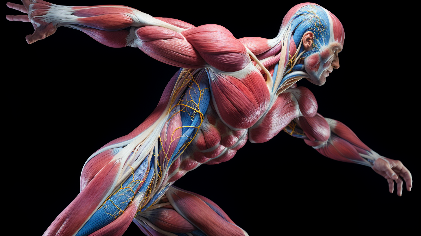
The functioning of muscles
What is the functioning of muscles? What are the processes in muscles that lead to muscle growth, and what is the role of nutrition and training at the cellular level? For the time and effort we spend in the gym and on nutrition, we often know relatively little about what exactly happens in the muscle. We train according to certain principles because we see that they work for others, but often cannot explain exactly why they work.You deliberately damage your muscles by training them, and during recovery, they grow through rest and nutrition.But if you then ask how they are damaged exactly, how they exactly grow, and why nutrition is important for this, most people don't get much further. Not strange in itself because all biological, physiological, and anatomical terms and processes can often be very complicated. However, it is worth taking the time to absorb this knowledge once in a while to get much more results from the countless hours of training and diet. So here are the most important points, (hopefully) explained in a way that most people can understand. Because otherwise, it would be a very extensive piece, I have divided it into several parts. In this first part, I will discuss the different types of muscles and their function and operation at the cellular level.
- In the second part, I will delve into how muscles grow, their application in training and nutrition, and the exact functioning of the different forms of muscle growth.
- In part three: The differences per person, body types endo-, meso-, and ectomorph
- In part four: The usefulness and function of warming up
- In part five: The usefulness and function of (various forms of) stretching
Different types of muscles
There are three different types of muscles in the body:- Smooth muscle tissue: Smooth muscles are muscles that we cannot consciously control. They are therefore also called involuntary muscles. Examples include the muscles of the gastrointestinal tract that move food through the digestive system and blood vessels (to regulate blood pressure and circulation) and airways that can become larger and smaller.
- Skeletal: This muscle tissue is called so because of the transverse stripes you see when you view this tissue under the microscope. Especially the so-called skeletal muscles (see below) consist of striated muscle tissue. They are connected to the somatic nervous system, which means that we can consciously control them.
- Cardiac muscle tissue: The tissue of the heart is striated but behaves like smooth muscle tissue in the sense that it is not consciously controlled.
Skeletal Muscles
Because I will delve deep later, I will further limit myself here to the muscles that interest us, the skeletal muscles. Skeletal muscles are so named because they connect two parts of the skeleton. Adult men and women consist on average of 42% and 36% skeletal muscles, respectively. In total, you have more than 660 skeletal muscles. The skeletal muscles are striated muscles that are controlled by the somatic nervous system, allowing us to consciously control them. The main functions of skeletal muscles are body movement and stabilization. Just like a hydraulic pump moves the arms of a crane, muscles do the same by becoming larger and smaller. They are attached in such a way that different parts of the body can move at the joint. Take, for example, the biceps brachii. This muscle allows you to bend your arm. The biceps attaches at the top to two places on the collarbone, runs over the upper arm (humerus) and the elbow, and is connected at the bottom to the radius. When the muscle becomes smaller, it pulls on the forearm, causing it to bend. Understanding the functions of the different skeletal muscles enables the development of exercises to strengthen and enlarge the muscles. By knowing that the biceps bend the arms, you know that you need to bend the arm to train the biceps. This may be a simple example that everyone says "Duhh," but I often get asked in the gym, "Do you know a good exercise for.....?" If you know the functions, you can actually come up with the exercises yourself, just as others have done for you. All fitness equipment is built based on this knowledge. Muscles can only pull! Muscles work by contracting together, causing them to shorten (contraction) and then relax and lengthen again. Like a rope, they can only generate force by pulling on something and not by pushing against it. During bench pressing, you push a weight up, but the chest muscle makes this possible by pulling the upper arm forward, inward, and upward. The arm bends as the biceps pull on the forearm (radius), and the triceps extend the arm by pulling on the ulna from the back.Origin and Insertion
The points where the muscle connects to bone are called origin and insertion. The origin, the point of origin, is often the non-moving part. In the given example, the collarbone is not moved, but the forearm is. In this case, the connections to the collarbone are the points of origin, and the forearm bone is the point of insertion. A muscle can have multiple points of origin and insertion. Another way to distinguish origin and insertion is the fact that the origin is often closest to the heart.Attachments
At the points where muscles attach to bone, they extend into a thin band of strong connective tissue made up of collagen and appear white. These are the attachments that transfer force to the bone, as happens in the example of the crane machine, for example, bolts connecting the hydraulic pump to the arm. Because these bands are thinner than the rest of the muscle and have poorer blood flow, they often form the weak link. Many bodybuilding injuries are often related to the attachment, but more on that in the article "Attachment Problems, Common Injury in Bodybuilders" (and/or separate articles on specific attachment problems of the chest, knee, biceps, or shoulders). The attachment(s) of the muscle closest to the heart is/are called the proximal attachment(s). The attachment(s) farthest from here is/are called the distal attachment(s). In the example of the biceps, the attachments to the collarbone are the proximal attachments, and the attachment to the forearm is the distal attachment.Different Skeletal Muscles, Shapes, Heads, and Fiber Direction.
Shapes of Muscles
Skeletal muscles come in different shapes. The most common are:- Fan-shaped
- Flat/belly-like
- Spindle-shaped
Heads of the Muscle
A muscle can have multiple heads. In the case of a multi-headed muscle, the muscle bundles extend into multiple attachments on the side of origin or insertion. The biceps brachii is a two-headed muscle (the name biceps refers to these two heads, just as triceps refers to three heads and quadriceps to four heads). If you look closely at the biceps image above, you'll see that at the top (the proximal part), it extends into two attachments. If you follow the attachments down to the muscle belly, you'll see that the muscle itself in the upper part is distinguished by a stripe. This stripe distinguishes the two heads to which the name refers. Just as various attachment points can provide different directions of movement, various heads can provide different directions of movement. Examples:- 1-headed muscle: Sartorius. The Sartorius is the longest muscle in the body and runs diagonally across the thigh.
- 2-headed muscle: Biceps brachii. As explained above, the biceps brachii has two heads with which it attaches to two points on the scapula. The two heads are distinguished by the names long head (caput longus) and short head (caput brevis).
- 3-headed muscle: Triceps brachii. The triceps brachii are responsible for extending the arms. The three heads are hardly visible to the eye. At the bottom, the muscle seems to split into two parts, the familiar horseshoe pattern. However, these two parts have one attachment to the ulna. The three heads (long, medial, and lateral) are found at the top. In the image above, these are clearly indicated in color.
- 4-headed muscle: The 4 muscles responsible for extending the leg share one attachment point to the kneecap and the shinbone. The quadriceps is formed by the vastus lateralis and vastus medius (respectively, outside and inside), the rectus femoris in the middle, and below it, the vastus intermedius. The sartorius is often considered one of the four heads of the quadriceps instead of the vastus intermedius, possibly because the intermedius is not visible in many images as it lies beneath the rectus femoris. However, the sartorius is indeed a separate, single-headed muscle. In the image on the left, the sartorius is the thin, long muscle running from the top right, diagonally over the quadriceps, to the bottom left.
Run your fitness business with more structure
- Planning, member management and coaching in one platform
- Built for coaches, studios and gyms
- Fewer separate tools, more overview
Fiber Direction
In addition to shape and the number of heads, muscles can also be classified based on the direction or directions in which the fibers (more on muscle fibers later) run. Next to you, you'll see various examples with names of muscles for illustration. An important difference lies between parallel and pennate muscles. The illustrated convergent and fusiform muscles are examples of parallel muscles; only the shape differs from, for example, the sartorius, which is why they have a modified name. If you take the length axis or axes (in the case of fan/convergent shape) from origin to insertion, you'll see that in these muscles, the fibers run parallel to these axis(es). If you look at the sartorius in the example next to it and draw a line between both ends (the attachments at origin and insertion), you'll see that the lines of the (bundles of) muscle fibers run parallel to this line. Then there are pennate muscles, named after the Latin for "feathered". In these muscles, the muscle fibers are perpendicular to the axis between origin and insertion. This allows more muscle fibers to be recruited, but over a smaller range of motion. Some hand muscles are an example of this. Next, there are bipennate muscles, where the fibers run in two directions from the axis, like the veins of a leaf or like a feather. The rectus femoris, the middle, superficial muscle of the quadriceps, is an example of this. When the attachment of a pennate muscle splits/branches into multiple attachments, we speak of a multipennate muscle, such as the deltoid, the large shoulder muscle. Finally, there are circular muscles whose fibers run in a circle. Examples of this are where food first enters and where it leaves your body. Knowledge of fiber direction is important because, like understanding the function of the muscle, it provides insight into how to train the muscle. Take the pectoralis major as an example, the large chest muscle (see example in images above). You can clearly see the fibers running horizontally and partly diagonally. If I now do bench presses on an inclined bench, so the arms are not perpendicular to the torso but at an angle of 30-45 degrees towards the head, then I use the upper fibers for this. These can thus be trained separately. Now you may want to focus on the inside of the chest by, for example, doing cable crossovers, but then differently than in this instructional video by actually crossing the hands (as the name of the exercise suggests) and concentrating on this part of the exercise. However , this doesn't make sense, at least not specifically for the inside of the chest. There are no separate fibers that only run on the inside of the chest, as there are fibers that only run on the top and bottom of the chest. So you train the muscle over the entire, in this case horizontal, path. This is important because you will often hear that you can do specific exercises for, for example, the inside of the chest or the bottom of the biceps (by focusing on the lower part of the muscle). This is not possible. For this, I wrote a separate article at the end of last year in case you want to start this discussion again in the gym. However, these exercises are good because they provide a different load and therefore variation in your training. The incorrect idea that you can train the inside of the chest or the bottom of the biceps comes in part from the saying that you only make a muscle grow in the part you train. However, this saying is not entirely correct and should read: "You only make a muscle grow in the part where you train the fibers".Naming of Muscles
Muscles can be named for various reasons. These can be named after:- Shape: Such as the shoulder muscles, deltoid, named after the triangular shape, and the trapezius after a trapezium
- Size: For example, Pectoralis major and minor, the large and small chest muscles.
- Number of heads: For example, bi-, tri-, and quadriceps.
- Direction of fibers in relation to the body's midline or the length axis of a bone or limb: For example, the rectus femoris, which runs in a straight line relative to the thigh (femur).
- Origin and insertion: In this case, the origin is mentioned first in the name. An example of this is the sternocleidomastoid ("breastbone-clavicle-nipple muscle"). This muscle has two points of origin, the sternum (breastbone) and clavicle (collarbone). Hence, the name begins with sterno (for sternum) and cleido (for clavicle). The insertion is not on the nipple as the Dutch name might suggest (too bad for the ladies looking for an alternative to a push-up bra) but on the mastoid process (process mastoideus), which is located on a projecting part of the temporal bone. This muscle is responsible for bending and rotating the neck.
- The action: Many muscles have, for example, "flexor" or "extensor" in their name. This refers to the bending (flexor) or stretching (extensor) action of the muscle.
Muscle Bundles and Fascia
Now that we've discussed the different types of skeletal muscles, let's take a look at the muscle itself and how it's structured. I'll try to strike a balance between providing comprehensive information and using understandable language, but it's quite challenging. I'll explain as many non-commonly known terms as possible, but unfortunately, I can't avoid making the following story somewhat complicated (the human body is complex). So far, I've often used the term muscle fiber, but what exactly is it? In the images above, you can see lines running through the muscle indicating the direction of the fibers. However, the muscle is not composed of a large number of individual fibers but of bundles containing muscle fibers. Just like the cables of a suspension bridge consist of multiple steel cables that are in turn made up of thinner cables. 1. Muscle bundle 2. Muscle fiber 3. Perimysium In the image on the right, you can see a (spindle-shaped) muscle. The muscle is composed of multiple muscle bundles that are collectively packed in fascia. (Deep) fascia is a plastic-like type of connective tissue that provides structural integrity throughout the body by keeping things in place and allowing muscles to slide over each other. The fascia that binds the entire muscle together is called the Epimysium (labeled "2. connective tissue membrane" in the image on the right). Within the muscle, the bundles of fibers themselves are also held together by fascia called Perimysium. Finally, each individual muscle fiber is also surrounded by fascia called Endomysium. All these different layers of fascia come together at the beginning and end of the muscle, forming the attachments.Examining the Muscle Fiber, Muscle Fiber = Muscle Cell
1. Myofibril 2. Blood vessel 3. Nucleus of the muscle fiber 4. The entire muscle fiber We've seen that a (skeletal) muscle, like the cables of a suspension bridge, is composed of multiple bundles, and these bundles are made up of fibers. If you were to cut through one of these fibers, you would see that it, too, is composed of multiple "cables". Before we proceed, it's important to understand that a muscle fiber is a cell, the smallest building block of the body. For those whose biology education didn't extend beyond school, they might imagine round transparent bubbles with a nucleus inside when thinking of cells. However, cells can take on many different shapes and contain varying numbers of nuclei and other contents. A muscle fiber is often not called a cell because it contains multiple nuclei and is formed by the fusion of multiple cells. However, since we'll be discussing processes at the cellular level, I'll often refer to a muscle cell (myocyte) for clarity. A muscle cell is a tubular cell that can be as long as 30 centimeters, as it can run the entire length of a muscle, with a thickness roughly that of a hair. The cell of a skeletal muscle originates from the fusion of multiple cells (myoblasts), thus containing multiple nuclei. The large number of nuclei per muscle cell allows it to hold more cytoplasm, contributing to its size. The "shell" of the muscle cell is formed by the plasma/cell membrane, which in the case of muscle cells is called the sarcolemma. The cytoplasm of the cell (for cells in general: cytoplasm) of a muscle is called sarcoplasm. The blood vessels provide oxygen, moisture, and nutrients and remove waste products. Additionally, the muscle cell contains mitochondria (singular: mitochondrion). These are the powerhouses of the muscle cell. A crucial function of mitochondria is to produce ATP (see box), adenosine triphosphate. ATP is the primary supplier of energy that enables muscle contraction. This brings us to the most important component of the muscle cell: the myofibrils, which are responsible for muscle contraction. I'll first delve into myofibrils in detail and then briefly revisit the function of the T-tubules and the sarcoplasmic reticulum.Myofibrils, Sarcomeres, and Filaments: The Tensioners of the Muscle
Most of the space within the cell is occupied by myofibrils, which ultimately cause muscle contraction. Myofibrils can be seen as long threads with the thickness of 1/100th of a hair that run through the entire length of the muscle fiber/cell. These threads are formed by chains of two different proteins, myosin and actin. In the image on the right, you can see a simplified depiction of how they work. Before I explain this mechanism, I'll first provide an explanation of myosin and actin. Myosin: In the middle, you can see the cylindrical myosin filament. Filament stands for "thread" or "fiber," and in the case of muscles, it refers to "protein thread." A myosin filament is a polymer (a molecule composed of multiple identical units) made up of myosin units with a head and a tail. These tails are intertwined so that the heads protrude on both outer sides. Actin: On the outer sides, you can see actin. Actin is also a polymer formed by two actin monomers (a monomer is a single chemical compound) forming a double chain. A myosin filament and the surrounding actin filaments together form one sarcomere, the functional unit of a muscle. Thus, a muscle is composed of bundles containing muscle fibers as thin as hair, which in turn contain dozens of myofibrils, each of which is made up of multiple chains of sarcomeres.No Binding of Myosin and Actin in the Relaxed State
Muscle contraction occurs as numerous myosin filaments along the entire length of a muscle cell grip onto actin filaments and pull them toward the center of the myosin filament. This gripping and releasing is a complex process. An actin filament has spaces at various locations where the heads of myosin filaments can bind (grip). This is a strong connection, also known as rigor.</p >Binding of Myosin and Actin by Calcium Ions
When you want to perform a movement, a stimulus from a motor nerve (a nerve that connects the central nervous system with the muscles) causes the release of the neurotransmitter (signaling molecule) acetylcholine in the synapse (the space where the transmission between two nerve cells takes place). The acetylcholine binds to receptors on the sarcolemma (the muscle cell membrane/"shell" around the muscle cell). Without delving too deeply into the polarity of the sarcolemma (it's complicated enough already), this causes depolarization of the sarcolemma with the release of calcium ions (Ca2+) as a result. These calcium ions bind to the troponin complex that is bound to the tropomyosin, which blocks the binding site for myosin. As a result, the calcium ions pull the troponin complex from its place, causing the tropomyosin bound to it to also be displaced, thereby exposing the binding site for myosin. The sarcoplasmic reticulum and the T-tubules ensure rapid propagation of the signal to act (T-tubules) and over (sarcoplasmic reticulum) the cell.Binding, Releasing, and Rebinding by ATP
The binding of myosin to actin described above does not occur just once but multiple times per contraction. The heads of the myosin filament grab, release, grab again, and thus pull the actin filament further toward the center. This process requires a lot of energy, which is derived from ATP that, as explained above, is produced in the mitochondria, the cell's powerhouses. When myosin is bound to actin, the myosin head then binds to ATP and magnesium. This breaks the connection with the actin filament, causing the heads to detach and rotate further outward. The myosin then fully binds with ATP. The enzyme ATP-ase hydrolyzes the ATP with the help of magnesium (splits by absorbing water), breaking it down into ADP (adenosine diphosphate) and phosphate. This releases energy that can be used to reattach the head (which is no longer bound to the split ATP) to the actin filament, but further along. This process pulls the actin further inward. ATP is also necessary for pumping back the calcium ions once the action is completed, so that the binding sites are covered again by tropomyosin. The image below shows how this process works with explanations of the various steps.The Importance of Magnesium
Magnesium is required for muscle relaxation. Magnesium also plays a role in ATP production. A deficiency in magnesium can therefore lead to cramps, muscle pain, and declining energy levels.Differences in Filament Numbers per Person and Strength Control
The strength per sarcomere is (under equal conditions such as available ATP) actually the same for everyone. However, the total number of filaments next to each other in one myofibril varies from person to person. For example, men have thicker muscles than women because, among other factors, they have a higher level of testosterone, which provides more filaments next to each other in a myofibril, making it thicker. A sarcomere as a motor unit utilizes all available strength at once. You can't give half throttle to a sarcomere. The extent to which you use strength depends on the number of motor units and which ones are used. The body will not use all sarcomeres, but only the necessary ones. More filaments = thicker myofibril = thicker muscle fiber = thicker muscle bundle = thicker muscle. Even when you relax, some sarcomeres are contracted. As mentioned at the beginning of this article, one of the functions is to maintain the shape of the bag of bones that your body is. This minimal tension to keep everything in place is called tone.Energy Sources of the Muscles
We have now seen that ATP is needed to contract and relax by grabbing and releasing actin. What happens when there is no more ATP? The heads of myosin cannot attach to ATP anymore and therefore remain attached to actin, keeping the muscle tense. This is exactly what happens in corpses. The so-called rigor mortis (stiffness of death) is caused by calcium ions leaking from the sarcoplasmic reticulum, allowing myosin to bind to actin. However, because no ATP is being produced, the heads cannot detach anymore. Additionally, in the absence of ATP, the calcium ions cannot be pumped back. Muscle cramps can also, in addition to a magnesium deficiency, be related to a shortage of ATP when, for example, not enough carbohydrates, proteins, and/or have been eaten. These nutrients can be stored (as glycogen, glycerol, and amino acids, respectively) in the body. When these substances are broken down by oxygen, energy is produced, which is stored as ATP.Three Energy Systems
The amount of ATP in the body is very limited and therefore needs to be continuously generated. The body has three systems to generate this energy. It is important whether the body receives more oxygen than it consumes at that moment. The maximum amount of oxygen you take in is often measured and expressed as VO2 max.Anaerobic Alactic Energy System / Phosphate System
-How?: Anaerobic means without the use of oxygen. You consume more oxygen than you take in. Alactic means without the formation of lactic acid. -Suitable for: Very intense, short exertion (10 -20 seconds) -Example: 100-meter sprint, weightlifting with heavy weights and few repetitions -Uses: Creatine phosphate + ADP -Produces: ATP -Special features: Your body consumes more oxygen than it takes in. This is not a problem as long as the duration of the activity is limited to about 10 seconds. If it lasts longer, strength decreases, and the body switches to the next energy system:Anaerobic Lactic Energy System
-How?: Without oxygen, but with the formation of lactic acid due to incomplete combustion of glucose (sugars) -Suitable for: Heavy exertion lasting 20 seconds to about 2 minutes, for example, running the 400 meters. -Uses: Glucose -> pyruvic acid -> lactic acid -Produces: ATP & lactic acid -Special features: Lactic acid causes "acidification" that can lead to cramps and must be broken down in the liver.Aerobic Energy System
-How?: Uses oxygen -Suitable for: Lighter exertion of longer duration -Uses: (Complete combustion of) glucose, fats, and protein (depending on duration, intensity, and available nutrients) by oxygen -Produces: CO2 + water + ATP -Special features: Carbohydrates are a more efficient energy source than fats because fats use more oxygen for their combustion. However, the carbohydrate supply is often much more limited than the fat supply. In the aerobic system, you could theoretically keep running indefinitely as long as someone gives you a sandwich or other form of carbohydrates every so often. The only reason this is not possible is due to the need for sleep (of which science still does not know exactly what its purpose is). Understanding the operation of energy systems is important to know what nutrients your body needs, when they are needed, in what quantities, and for what purpose.Curious how FITsociety fits your business?
See how intake, training, payments and communication come together.
Start a free trial and explore the workflow at your own pace.